39 microscope images with labels
Compound Microscope - Diagram (Parts labelled), Principle and Uses Compound Microscope - Diagram (Parts labelled), Principle and Uses Compound Microscope As the name suggests, a compound microscope uses a combination of lenses coupled with an artificial light source to magnify an object at various zoom levels to study the object. A compound microscope: Is used to view samples that are not visible to the naked eye Microscope Images Labeled | Virtual Anatomy Lab VAL © 2023 by Yvonne Szymanski, Lisa Dubuc, & Bob Bucella Proudly created with Wix.com Wix.com
Compound Microscope Parts - Labeled Diagram and their Functions - Rs ... The eyepiece (or ocular lens) is the lens part at the top of a microscope that the viewer looks through. The standard eyepiece has a magnification of 10x. You may exchange with an optional eyepiece ranging from 5x - 30x. [In this figure] The structure inside an eyepiece. The current design of the eyepiece is no longer a single convex lens.

Microscope images with labels
Labeling the Parts of the Microscope Labeling the Parts of the Microscope This activity has been designed for use in homes and schools. Each microscope layout (both blank and the version with answers) are available as PDF downloads. You can view a more in-depth review of each part of the microscope here. Download the Label the Parts of the Microscope PDF printable version here. Histology and Microscope Slide Labels Microscope Slide Labels These specialty Microscope Slide Labels and matching End Labels are available in standard (thin) or pathology (tissue high) thickness, and square or round corner (RC). Permanent adhesive holds labels in place during use and long-term storage. Sheet Form Size is 5¼" x 8". Prices are per thousand labels Slide Label Microscope picture label Flashcards | Quizlet Microscope picture label Flashcards | Quizlet Microscope picture label STUDY Flashcards Learn Write Spell Test PLAY Match Gravity Created by kfire Terms in this set (12) Arm What is the part labelled C? Base What is the part labelled D? Body tube What is the part labelled B? Ocular lens What is the part labelled A? Illuminator
Microscope images with labels. Microscope Parts, Function, & Labeled Diagram - slidingmotion Microscope parts labeled diagram gives us all the information about its parts and their position in the microscope. Microscope Parts Labeled Diagram The principle of the Microscope gives you an exact reason to use it. It works on the 3 principles. Magnification Resolving Power Numerical Aperture. Parts of Microscope Head Base Arm Eyepiece Lens 26+ Picture Of A Microscope With Label PNG 26+ Picture Of A Microscope With Label PNG. Microscopes are specially created to magnify the image of the subject being studied. Students label the parts of the microscope in this photo of a basic laboratory light microscope. Microscope Drawing And Label at GetDrawings | Free download from getdrawings.com I searched for this on bing.com/images. Compound Microscope Parts, Functions, and Labeled Diagram Compound Microscope Definitions for Labels. Eyepiece (ocular lens) with or without Pointer: The part that is looked through at the top of the compound microscope. Eyepieces typically have a magnification between 5x & 30x. Monocular or Binocular Head: Structural support that holds & connects the eyepieces to the objective lenses. What are the parts of a microscope labeled? - SidmartinBio Label the microscope. Use this interactive to identify and label the main parts of a microscope. Drag and drop the text labels onto the microscope diagram. ... Which is the body tube of a microscope? Image 2: The body tube part of a microscope is where the ray of light is bent to allow the object being viewed to enlarge by the scope. Picture ...
Parts of a microscope with functions and labeled diagram Optical parts of a microscope and their functions The optical parts of the microscope are used to view, magnify, and produce an image from a specimen placed on a slide. These parts include: Eyepiece - also known as the ocular. This is the part used to look through the microscope. Its found at the top of the microscope. Parts of a Simple Microscope - Labeled (with diagrams) image 2: A simple microscope commonly used by students for studying minute objects. image source: imimg.com. picture 3: It is the latest design of a simple microscope - advanced features than the conventional simple microscopes. ... image 5: A modern simple microscope with the different parts labeled. image source: laboratoryinfo.com. The ... 300+ Free Microscope & Laboratory Images - Pixabay Upload 399 Free images of Microscope Related Images: laboratory science bacteria research scientist lab biology chemistry medical Find your perfect microscope image. Free pictures to download and use in your next project. 399 Free images of Microscope / 4‹ › Amazing 27 Things Under The Microscope With Diagrams Skeletal muscle under the microscope 40X magnification 100X magnification 400X magnification 20. Skin under the microscope 21. Snowflake under the microscope 22. Sperm under the microscope Direct observation Observation after staining 23. Spirogyra under the microscope 24. Virus under the microscope Fluorescence microscope
Parts of the Microscope with Labeling (also Free Printouts) Microscopes are specially created to magnify the image of the subject being studied. This exercise is created to be used in homes and schools. the microscope layout, including the blank and answered versions are available as pdf downloads. Click to Download : Label the Parts of the Microscope (A4) PDF print version. Label the microscope — Science Learning Hub All microscopes share features in common. In this interactive, you can label the different parts of a microscope. Use this with the Microscope parts activity to help students identify and label the main parts of a microscope and then describe their functions. Drag and drop the text labels onto the microscope diagram. Microscope Types (with labeled diagrams) and Functions This is an advanced microscope that has specific application in viewing, observing and measuring the optical thickness and phase of completely transparent specimens and objects. A tiny interferometer is used and a specimen is placed on beam path of it. This path is split and then rejoined to create two superimposed images of the specimen in focus. Microscope Parts and Functions Microscope Parts and Functions With Labeled Diagram and Functions How does a Compound Microscope Work?. Before exploring microscope parts and functions, you should probably understand that the compound light microscope is more complicated than just a microscope with more than one lens.. First, the purpose of a microscope is to magnify a small object or to magnify the fine details of a larger ...
Labeling a Compound Microscope - PurposeGames
Microscope Labeled Pictures, Images and Stock Photos Browse 48 microscope labeled stock photos and images available, or start a new search to explore more stock photos and images. Newest results Fluorescent Imaging immunofluorescence of cancer cells growing... Plant Tissue Systems vector illustration. Labeled biology... Microscope diagram vector illustration. Labeled zoom instrument...

Label the microscope Quiz
Compound Microscope with labels Stock Vector | Adobe Stock Download Compound Microscope with labels Stock Vector and explore similar vectors at Adobe Stock. Adobe Stock. Photos Illustrations Vectors Videos Audio Templates Free Premium Editorial Fonts. ... Get 10 free Adobe Stock images. Start now. Get 10 free images. Unlock 200M+ assets in our full collection.
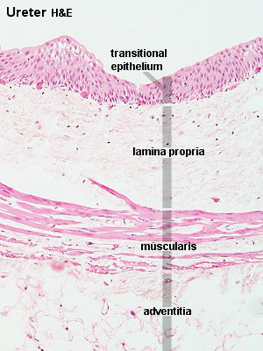
ANAT2511 Urinary System - Embryology
Microscope Drawing And Label - Painting Valley Are you looking for the best images of Microscope Drawing And Label? Here you are! We collected 33+ Microscope Drawing And Label paintings in our online museum of paintings - PaintingValley.com. ADVERTISEMENT LIMITED OFFER: Get 10 free Shutterstock images - PICK10FREE label microscope diagram compound parts light labeling functions microscopic
Zoeken in galerij
Microscope, Microscope Parts, Labeled Diagram, and Functions Microscope, Microscope Parts, Labeled Diagram, and Functions What is Microscope? A microscope is a laboratory instrument used to examine objects that are too small to be seen by the naked eye. It is derived from Ancient Greek words and composed of mikrós, "small" and skopeîn,"to look" or "see".

31 Picture Of A Microscope To Label - Labels For Your Ideas
A Study of the Microscope and its Functions With a Labeled Diagram A Study of the Microscope and its Functions With a Labeled Diagram To better understand the structure and function of a microscope, we need to take a look at the labeled microscope diagrams of the compound and electron microscope. These diagrams clearly explain the functioning of the microscopes along with their respective parts.

Microscope With Labels free vector | Download it now!
Polarizing Microscope Image Gallery - Leica Microsystems The position of the optical axis can be clearly determined with circular polarization. Right: Conoscopic image of the same calcite sample with linear polarized light. The calcite section is perpendicular to the optical axis. Images recorded with a DM4 P microscope using transmitted light, conoscopy, 63x N Plan objective, and polarizers.

Labeling The Microscope - PurposeGames
Microscope With Labels clip art | Microscope parts, Scientific method ... Jul 3, 2012 - Download Clker's Microscope With Labels clip art and related images now. Multiple sizes and related images are all free on Clker.com.

labels of a compound microscope microscope boxed - Top Label Maker
Microscope labels | Bill Todt Microscope labels. Unlabelled image: Lab Index: 101 Class Page: Identify the following parts of the microscope: Eyepieces Observation tube Nosepiece Objectives Stand Specimen holder Mechanical stage Stage controls Condenser Iris diaphragm Coarse focus Fine focus

Microscopic Structure of Bone | ClipArt ETC
PDF Label parts of the Microscope Label parts of the Microscope: . Created Date: 20150715115425Z
Search in gallery
Microscope Labeling Game - PurposeGames.com About this Quiz. This is an online quiz called Microscope Labeling Game. There is a printable worksheet available for download here so you can take the quiz with pen and paper. This quiz has tags. Click on the tags below to find other quizzes on the same subject. Science.

Microscope Picture To Label - Micropedia
Parts of Stereo Microscope (Dissecting microscope) - labeled diagram ... Stereo microscopes (also called Dissecting microscope) are branched out from other light microscopes for the application of viewing "3D" objects. These include substantial specimens, such as insects, feathers, leaves, rocks, sand grains, gems, coins, and stamps, etc. Functionally, a stereo microscope is like a powerful magnifying glass.
Quia - Protist Vocabulary
Microscope picture label Flashcards | Quizlet Microscope picture label Flashcards | Quizlet Microscope picture label STUDY Flashcards Learn Write Spell Test PLAY Match Gravity Created by kfire Terms in this set (12) Arm What is the part labelled C? Base What is the part labelled D? Body tube What is the part labelled B? Ocular lens What is the part labelled A? Illuminator

Haversian System
Histology and Microscope Slide Labels Microscope Slide Labels These specialty Microscope Slide Labels and matching End Labels are available in standard (thin) or pathology (tissue high) thickness, and square or round corner (RC). Permanent adhesive holds labels in place during use and long-term storage. Sheet Form Size is 5¼" x 8". Prices are per thousand labels Slide Label
Search in gallery
Labeling the Parts of the Microscope Labeling the Parts of the Microscope This activity has been designed for use in homes and schools. Each microscope layout (both blank and the version with answers) are available as PDF downloads. You can view a more in-depth review of each part of the microscope here. Download the Label the Parts of the Microscope PDF printable version here.

Microscope Journal – The Indigo Teacher

Lysosomes Dr.Jastrow's electron microscopic atlas
Label a microscope - Teaching resources
Post a Comment for "39 microscope images with labels"